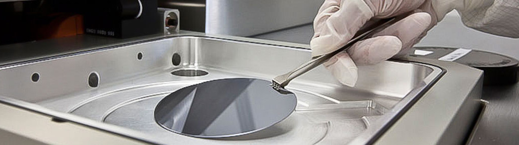
Integrated Nanosystems Research Facility


Microfluidic Biochip
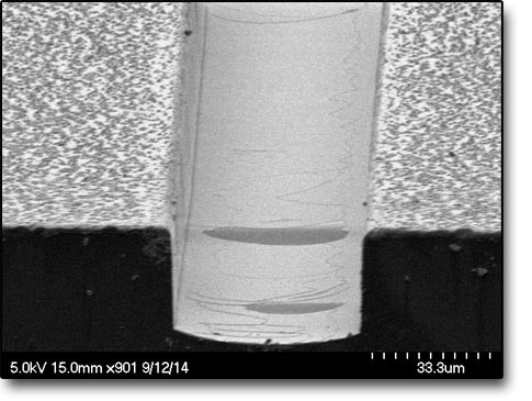 This is a scanning electron microscope (SEM) image of a microfluidic biochip on a fused silica substrate. The image shows the nickel hard mask used during dry etching.
Equipment used:
- E-beam 1 evaporator to deposit a seed layer on the fused silica substrate.
- Laurell Spinner to spin photoresist.
- KS MA6 Mask Aligner to UV expose and pattern the fused silica substrate.
- Electroplating station to plate the nickel hard mask.
- SPTS Glass Etcher to dry etch the microfluidic channels.
- Dektak XT or Dektak 3 profilometers to measure channel’s etch depth.
- Optical microscope to inspect the completed biochip.
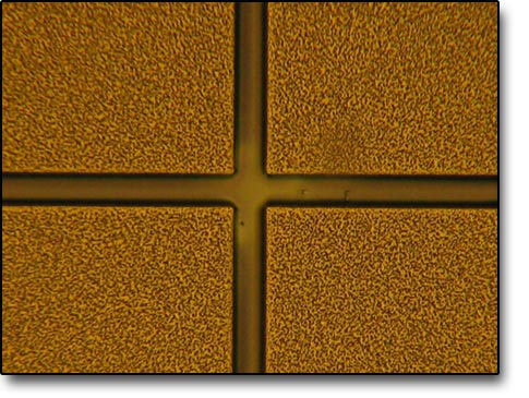 This is an optical microscope image of a microfluidic biochip on a fused silica substrate. The image shows the nickel hard mask used during dry etching.
Equipment used:
- E-beam 1 evaporator to deposit a seed layer on the fused silica substrate.
- Laurell Spinner to spin photoresist.
- KS MA6 Mask Aligner to UV expose and pattern the fused silica substrate.
- Electroplating station to plate the nickel hard mask.
- SPTS Glass Etcher to dry etch the microfluidic channels.
- Dektak XT or Dektak 3 profilometers to measure channel’s etch depth.
- Optical microscope to inspect the completed biochip.